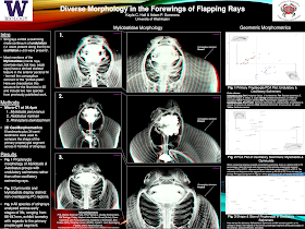
This week, I have a guest post from Kayla Hall, who created this poster. Click to enlarge!
I reached out to Kayla because all of her central images are in 3-D!
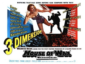
These images can be viewed in 3-D using the red-green glasses, which is the same technique that was used by filmmakers in the 1950s for movies like House of Wax and Creature from the Black Lagoon. Even Alfred Hitchock made a 3-D movie: Dial “M” for Murder. Interest in 3-D movies was high for a few years, then petered out.
This red/green 3-D image is technically called an anaglyph. I’ve seen this technique used rarely, but consistently, at poster sessions through the years, but never had reason to make one myself. I wanted to know how it was done.
First of all, why? Kayla wrote (lightly edited):
I had a few reasons for choosing to display these specimens in 3-D:
Firstly, our previous publication characterized a skeletal structure in the fins of one family of stingrays, but that project solely used 2-D radiography. This was the first time we’ve been able to see this morphology in 3-D space and quantify all of the other aspects, such as thickness and curvature depth (for muscle attachment) of the primary cartilages across families.
Second, most of my work has used specimens from museum collections. Computerized tomography (CT) scanning allows us to gather all of the anatomical and morphological information without destructively sampling rare specimens. In fact, two of the stingrays displayed on the poster are actually new species to us, not previously included in our publication, so this was a first for visualizing and characterizing their anatomy. Scanning and viewing the Myliobatis specimen (#1) in 3-D space allowed us to add this new species to the list of individuals that lack the pectoral fin framework, as this is the only genus to exhibit variation in the presence/absence of this trait across species.
So that’s the why, but what about the how?
We used the Bruker micro-CT scanner, reconstructed the CT images using the program NRecon, and finally visualized the reconstruction in CTVox. CTVox is also the program I used to generate the 3-D images. They have a “Stereo viewing” button that converts the reconstruction into a 3-D image viewable with standard red-blue glasses.
You are not able to tweak the red-blue hues, but you can always toggle with the original histogram settings that produce the 3-D reconstruction to alter the color contrast.
I bought a multi-pack of basic red and blue glasses from an online retailer. (Search “anaglyph 3-D glasses” or just “3-D glasses.”- ZF)
Printing on matte paper works best for visualizing the 3-D work.
Easy once you know how!
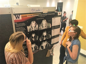
And here Kayla shows off her results to Kelsi Rutledge and Jules Chabain.
You can produce the 3-D effect with just a high-quality graphic editor. In brief:
- Get two images. You either need to take or find two pictures from slightly different viewpoints, or you need to edit an image to mimic the effect.
- Merge the photos.
- Remove the red from one image.
- Remove green and/or blue from one image.
- Crop the edges where they don’t overlap.
If you do this to a single image, however, you are creating an illusion. It isn’t “real data” in the sense that am image generated using a CT scan or two actual photographs. Be sure to label images with appropriate disclaimers!
External links
Diverse morphology in the forewings of flapping rays
How To Create Anaglyph 3D Images That Really Work!


When is your baby👶 due? How big is your baby👶 this week? Use seotoolsrack.com Pregnancy Due Date Calculator to estimate your baby's👶 due date based on the first day of your last period, the date you conceived and other methods. Your health care provider uses the number of weeks since your last menstrual period to describe how far along you are in your pregnancy.
ReplyDeletehttps://www.seotoolsrack.com/pregnancy-due-date-calculator